Locomotion and Movement:
Muscle: Muscle, which constitute about 40-50 percent of the body weight is of mesodermal origin.
Based on location, muscle is divided into: skeletal, smooth and cardiac muscles.
Comparison table of muscles are given below:
Based on location, muscle is divided into: skeletal, smooth and cardiac muscles.
Comparison table of muscles are given below:
|
|
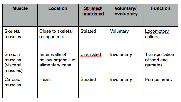
|
Click here for video for types of muscles.
Skeletal Muscle:
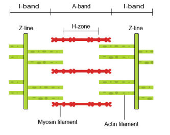
Each skeletal muscle is made up of a number of muscle bundles or fascicles connected by a sheath of connective tissue called fascia. And each muscle bundle is made up of a number of muscle fibres. Each muscle fibre is enclosed by sarcolemma enclosing sarcoplasm, which contains many nuclei and endoplasmic reticulum (store house of Calcium ion). Sarcoplasm contains a large number of parallelly arranged filaments called myofilaments or myofibrils, which contain light and dark band on it.
The light band (I-band or Isotropic band) contains actin (thin filament) and the dark band (A-band or Anisotropic band) contains myosin. actins are held together in the center by an elastic fibre called “Z” line and myosin filaments are held together in the middle by a thin fibrous membrane called “M” line. The portion of myofibrils between two successive “Z” line is the called sarcomere. In resting phase ends of actin filaments partially overlap the ends of myosin filaments.
Video for Structure of Skeletal Muscle.
|
|
|
Structure of Actin and Myosin:
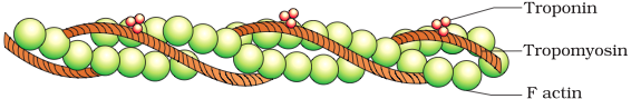
Actin: An actin is made up of two “F” (filamentous) actin, polymer of monomeric “G” (globular) protein, helically wounded around each other. Two filaments of another protein, tropomyosin, on which complex protein troponin is distributed at regular intervals, also run close to the two “F” actins. During resting state, subunit of troponin masks the active binding site for the myosin on the actin filaments.
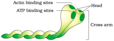
Myosin: Each actin filament is made up of monemeric protein meromyosin. Meromyosin has two parts: a globular head and a short arm-also called heacy meromyosin (HMM), cross arm- and a tail also called light meromyosin (LMM). The globular head is an active ATPase and has active binding sites for ATP and active sites for actin.
Myosin: Each actin filament is made up of monemeric protein meromyosin. Meromyosin has two parts: a globular head and a short arm-also called heacy meromyosin (HMM), cross arm- and a tail also called light meromyosin (LMM). The globular head is an active ATPase and has active binding sites for ATP and active sites for actin.
An actin filament (thin filament).
|
Monomer (meromyosin) of an myosin filament.
|
Mechanism of Muscle Contraction (sliding filament theory):
|
|
Muscle contraction is initiated by a signal sent by the central nervous system (CNS) through motor neuron. When the neural signal reaches the neuromuscular junction or motor-end plate, neurotransmitter (Acetyl choline) is released which generates an action potential in the sarcolemma that spreads throughout the muscle fibre. The action potential will cause the release of calcium ion from endoplamic reticulum that binds with subunit of troponin on actin unmasking the active binding site for myosin.
Myosin head will now use the energy from the hydrolysis of ATP to bind with the exposed active site on actin to form a cross bridge, which pulls the actin filaments towards the center of “A” band causing the shortening of sarcomere i.e. contraction. |
Myosin will go back to its relaxed state after releasing ADP and iP and again bind with ATP to repeat the process until the calcium ions pumped backed to sarcoplasmic cisternae resulting in making of active site on actin filaments after which actin filaments will move back to its original state i.e.relaxation.
Therefore during contraction, “I” bands gets reduced while “A” bands retain the length.
Repeated activation of the muscles will lead to fatigue because of accumulation of lactic acid due to anaeobic breakdown of glycogen.
Video for Sliding Filament Theory.
Therefore during contraction, “I” bands gets reduced while “A” bands retain the length.
Repeated activation of the muscles will lead to fatigue because of accumulation of lactic acid due to anaeobic breakdown of glycogen.
Video for Sliding Filament Theory.
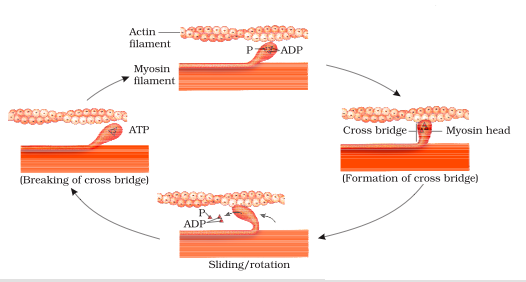
Diagram showing show cross bridge formation.
|
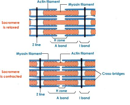
Diagram showing relaxed and contracted sarcomere.
|
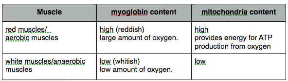
- Skeletal muscle can be divided into: red and white muscles.
The Human Skeletal System
- bone has very hard matrix due to calcium salts while cartilage has pliable matrix due to chondroitin salts.
- Human skeletal system consists of 206 bones.
- Skeletal system consists of 1.Axial skeletal 2.Appendicular skeletal.
1. AXIAL SKELETAL: (80 bones)
- Axial skeletal system is comprised of (i)skull, (ii) vertebrae, (iii) sternum, and (iv) ribs.
|
(i) Skull: (22 bones)
|
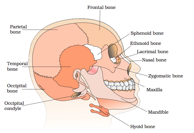
Diagrammatic view of human skull.
|
|
(ii) Vertebral column: (26 serially arranged units called vertebrae which are placed dorsally).
|
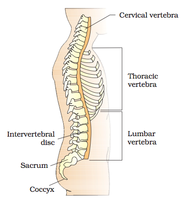
Diagrammatic view of vertebral column.
|
|
(iii) Sternum: sternum is a flat bone on the ventral mid-line of thorax.
(iv) Ribs: (12 pairs of ribs):
Video for Axial Skeletal. |
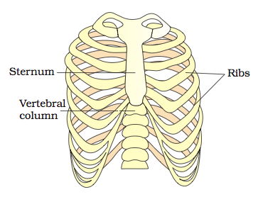
Diagram showing Ribs and rib cage.
|
2. APPENDICULAR SKELETAL: (limbs along with girdles constitute the appendicular skeleton).
(i) Limb:
(ii) Girdles: (pectoral and pelvic girdle):
Video for Appendicular Skeletal.
(i) Limb:
- Bones of hand are: Humerus, Radius, Ulna, Carpals (8 bones), Metacarpals (5 bones), Phalenges (14 bones).
- bones of leg are Femur (thigh bone- longest bone), Tibia, Fibula, Patella, Tarsals ( 7 bones), Meta tarsals (5 bones), and Phalenges (14 bones).
(ii) Girdles: (pectoral and pelvic girdle):
- Pectoral girdle:
- Formed by scapula and clavicle (collar bone).
- Clavicle articulates with scapula at acromion.
- below the acromion is a depression called Glenoid cavity to which humerus articulates to form shoulder joint.
- Pelvic girdle:
- pelvic girdle consists of two coxal bones.
- each coxal bone is made comprised of ilium, ischium, and pubis.
- at the point of fusion of these three bones is a cavity called acetabulum to which Femur articulates.
- Two coxal bones meet ventrally to form pubic symphysis.
Video for Appendicular Skeletal.
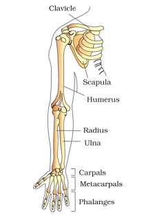
right upper limb and upper arm.
|
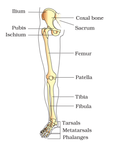
right pectoral girdle and lower limb.
|
|
HTML Comment Box is loading comments...
|
|
