Neural Control and Coordination
Neural System:
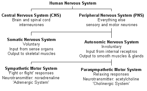
Neural system in animal is composed of highly specialised cells called neurons that can detect, receive and transmit different kinds of stimuli.
Human Neural System:
Summary of Human neural system is given below:
Neural system in animal is composed of highly specialised cells called neurons that can detect, receive and transmit different kinds of stimuli.
Human Neural System:
Summary of Human neural system is given below:
|
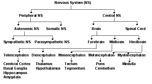
|
Click on the table to enlarge.
|
Click on the table to enlarge.
|
The nerves of PNS are of two types: (i) afferent fibres and (ii) efferent fibres.
- afferent fibres: transmit impulses from tissues/organs to the CNS and
- efferent fibres: transmit impulses from CNS to tissues/organs.
Structure of Neuron:
Neuron is the structural and functional unit of neural system. It consists of dendrite, cell body and axon. Cell body, also called cyton or soma, transmits the signal from dendrites to axon. The cytoplasm of cyton contains cell organelles and granular bodies called Nissl’s franules.
Short fibres that project out of cell body are called dendrites. The long fibre that emerges out of cyton is called axon. Bulb-like structure, also known as synaptic knob, present at the distal end axon contains neurotransmitter. Axon is divided into myelinated and non-myelinated axon fibre. Axons that are enveloped by Schwann cells that form myelin sheaths around the axons are called myelinated axon fibres, and are found in spinal and cranial nerves. The gaps between two myelin sheaths are called the nodes of Ranvier. Axon fibres that are enveloped by schwann cells that does not form myelin sheaths around the axons are called non-myelinated axon fibres, and are found in autonomous and somatic neural system.
Diagrammatic representation for structure of neuron is shown in Figure 1.
Based on the number of axon and dendrites, the neurons are divided into multipolar, bipolar and unipolar. Difference between multipolar, bipolar, and unipolar neuron is shown is Table 1.
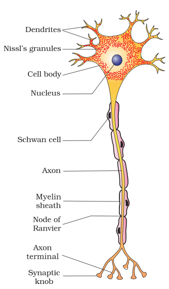
Figure 1.
Structure of a neuron.
Source: CBSE textbook
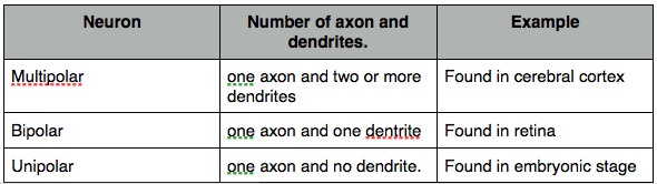
Table 1.
Comparison of multipolar, bipolar and unipolar neurons.
Source: CBSE textbook
Generation and Transmission of Nerve impulse:
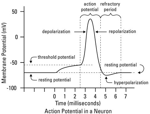
Transmission of an impulse
Types of Synapses:
Impulse is transmitted from one neuron to the other through a junction called synapses.
There are two types of synapses : electrical synapses and chemical synapses.
Differences between electrical and chemical synapses are given below:
There are two types of synapses : electrical synapses and chemical synapses.
Differences between electrical and chemical synapses are given below:
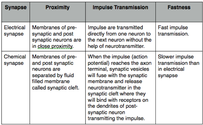
Differences between electrical and chemical synapse
The Structure of Brain:
Brain is protected by the skull. The brain is covered by three layers of cranial meninges consisting of dura mater (outer layer), arachnoid (middle layer) and pia mater (outer layer).
The brain is divided into three parts: (a) forebrain, (b) midbrain and (c) hindbrain.
(a) Forebrain: consists of (i) cerebrum, (ii) thalamus and (iii) hypothalamus.
(i) Cerebrum or the cerebral cortex: is the largest part of the brain and is divided into left and right hemisphere connected by a tract of nerve fibres called corpus callosum. Cerebral cortex is divided into four sections: frontal lobe, parietal lobe, occipital lobe and temporal lobe. Frontal lobe is associated with reasoning, part of speech, emotions and problem solving. Parietal lobe is associated with movement, orientation and perception of speech. Occipital lobe is associated with visual perception and processing. Temporal lobe is associated with perception and processing of auditory stimulus and memory.
(ii) Thalamus: is located below the fore brain. Thalamus is associated with coordination of sensory and motor signalling.
(iii) Hypothalamus: is located at the base of thalamus. Hypothalamus is associated with controlling body temperature. Hypothalamus contains some neurosecretory cells that secrete hypothalamic hormones.
Limbic system: made up of thalamus, hypothalamus, amygdala and hippocampus is located deep within the cerebrum. Limbic lobe is involved in the expression of emotional reactions and regulation of sexual behavior.
(b) Midbrain (mesencephalon): is located between the forebrain and hindbrain and consists of tectum and tegmentum. A canal called cerebral aqueduct passes through the mid brain. Corpus quadrigamina is four round swellings found at the dorsal portion of the midbrain.
(c) Hindbrain: consists of (i) pons, (ii) crerebellum and (iii) medulla oblongata.
(i) Pons: is involved in motor control and sensory analysis.
(ii) Cerebellum controls body movement and posture.
(iii)Medulla is responsible for controlling vital involuntary activities like respiration, cardiovascular activities and gastric secretions.
Midbrain and Hindbrain together form Brain stem.
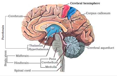
Structure of brain. Souce:cbse
The Structure of an Eye:
Structure of Eye:
The wall of eye consists of three layers: sclera, choroid and retina.
Sclera: is the outermost layer made up of dense connective tissue. The anterior potion of sclera is call cornea.
Choroid: is the middle layer that contains blood vessels. Anterior thick portion of choroid is called the ciliary body, which continues forward to form opaque structure called iris. The aperture surrounded by iris is called the pupil. Transparent eye lens held by ligament is also attached to ciliary body.
Retina: is the innermost layer that contains three layers of cells: ganglion (innermost), bipolar (middle) and photoreceptor cells (outermost). There are two types of photoreceptor cells: rods and cones. The cones perceive daylight vision while rods perceive vision in night. The photopigment in the rod is called the rhodopsin or the visual-purple. And rhodopsin is composed of opsin (a protein) and retinal (an aldehyde of vitamin A). There are three types of cones that contain their own characteristic pigments that respond to red, green and blue lights.
Region where the optical nerves leave the eye ball is devoid of any photoreceptor cells are called the blind spot. Macula lutea, the yellow portion of retina at the posterior pole, has a central pit, which contains only cones, called fovea where the resolution is greatest.
The space between the cornea and the lens is called the aqueous chamber that contains aqueous humor. The space between the lens and the retina is called the vitreous chamber that contains vitreous humor.
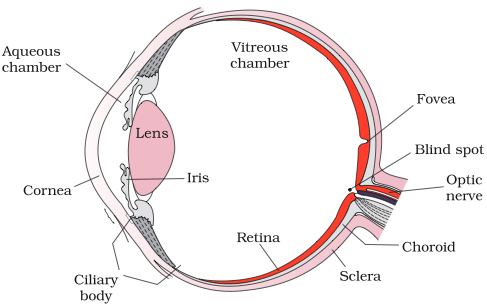
The structure of an eye. Source: cbse
Mechanism of Vision:
The light focused on the retina will cause the dissociation of retinal from opsin resulting in changes in the structure of opsin, which causes changes in membrane permiability in photoreceptor cells creating an impulse (action potential). This impulse (action potential) is passed on to the ganglion cells through the bipolar cells. The optic nerves will transmit the impulse to the visual cortex area of the brain, where the impulse is analyzed.
The light focused on the retina will cause the dissociation of retinal from opsin resulting in changes in the structure of opsin, which causes changes in membrane permiability in photoreceptor cells creating an impulse (action potential). This impulse (action potential) is passed on to the ganglion cells through the bipolar cells. The optic nerves will transmit the impulse to the visual cortex area of the brain, where the impulse is analyzed.
Ear and Hearing Mechanism:
HTML Comment Box is loading comments...
|
|
